
Editorial note: This English page is based on an educational article by IPD USA. The content has been translated, condensed and terminologically aligned with this website, while absolute marketing statements have been replaced with a more practical interpretation for the dental practice and laboratory.
What defines a digital workflow
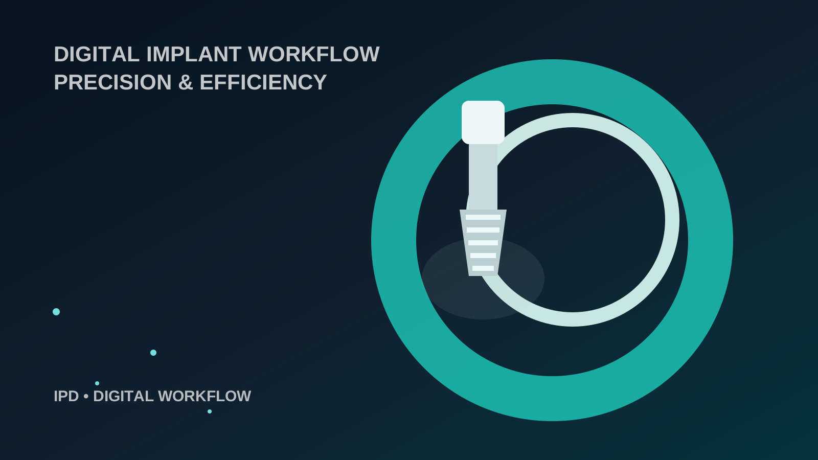
Digital implant prosthodontics is not a single step or a single device. It is a continuous data chain from the clinical scan through geometry identification to design and manufacturing. Each stage inherits information from the previous stage and can also add its own deviation to the final result.
The practical value of digitalisation is not speed alone. Its main advantage is the ability to standardise, repeat and verify individual steps. Where conventional impressions and stone models introduced material deformation, the digital pathway introduces different variables: scan quality, scan-body seating, mesh-to-library matching and manufacturing accuracy.
Where accuracy is actually created
Practical protocol for clinic and laboratory
- Verify complete scan-body seating visually and, where clinically appropriate, radiographically.
- Keep the connection area clearly readable by the scanner, free of interfering fluids and overlapping soft tissue.
- In the laboratory, verify not only the implant brand but also the platform, scanning level and exact scan-body reference.
- When a discrepancy occurs, do not automatically blame one component. Check the entire chain from scanning and library selection through to manufacturing.
How to read this article
The source text from IPD USA is educational and product-oriented material, not an independent clinical study. Marketing claims have therefore been moderated here, with the focus placed on continuity and verifiability throughout the digital workflow.
Where to continue on the website
Original source
Rafael Saucedo: Digital implant workflow: precision and efficiency, IPD USA, 2026.